MAMA Center for Reproductive Medicine
We’ve been helping couples with infertility problems since 1999. MAMA Clinic combines broad experience in IVF with cutting edge scientific research and individual approach to each patient. The highest pregnancy rate, shown by MAMA Clinic over the years, is the result of applying progressive Assisted Reproductive Techniques (such as In Vitro Fertilization and Intrauterine Insemination) performed with high professional standards.
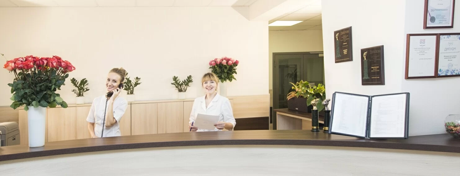
MAMA Clinic offers full range of IVF cycle procedures, which means our patients can have every test run and every procedure performed in one place, from standard set of initial survey to the final protocol.
MAMA Clinic holds European Standard Certificate and operates under Russian Federation regulations.
Phone: +7 495 921 34 26
WhatsApp: +7 926 206 35 17
Our call number operates from Monday to Friday in working hours from 9 a.m. to 6 p.m. (Moscow time)
Address: Russia, Moscow: 32, Raskovoi street
Nearest subway stations are Dinamo, Savyolovskaya
MAMA Clinic for Reproductive Medicine has been helping couples with fertility problems to achieve their dream of childbirth since 1999. MAMA Clinic combines broad experience in IVF with innovative scientific research and individual approach to each patient. The highest pregnancy rate, presented by MAMA Clinic over the years, is the result of applying the most progressive IVF treatment methods (such as Assisted Reproduction Technology and Intrauterine Insemination).
MAMA Clinic performs full range of ART cycle procedures including preliminary diagnostics to determine the reason of infertility followed by effective pre-insemination treatment and the EVF cycle itself. We take special pride in our pregnancy rates which are significantly higher than average in Russia. In 2013 the pregnancy rate shows 44.3% effectiveness (compared to average 37.5% estimate over the country), which is also considered to be very high across the Europe.
MAMA Clinic holds European Standard Certificate and operates under Russian Federation regulations.
Phone: +7 495 921 34 26
WhatsApp: +7 926 206 35 17
Our call number operates from Monday to Friday in working hours from 9 a.m. to 6 p.m. (Moscow time)
Address: Russia, Moscow: 32, Raskovoi street
Nearest subway stations are Dinamo, Savyolovskaya